Maxillary Tuberosity And Hamular Notch . The notch marks where the posterior border of the tuberosity (2) joins the medial (3) and lateral (4) plates of the pterygoid process. It is the posterior limit of the maxilla. The hamular notch is a loose connective tissue, about 2 mm wide, located between the maxillary tuberosity and the. The hamular notch is a groove between the maxillary tuberosity and the hamular process; Anatomical landmarks such as the retromolar pads, external oblique, mentalis muscle, frenum attachments, mylohyoid ridge, tuberosities, hamular notches, incisive papilla, labial sulcus, and buccal vestibule are critical and must be captured in the positive (and easily read in the negative) aspect of any edentulous impression. The hamular notch is critical to the design of the maxillary denture. The maxillary wax rim can be based on anatomic norms and is usually 22 mm high in the midline and 5 mm high in the hamular notch. Improper molding of this area could lead to soreness and loss of retention.
from www.slideserve.com
The hamular notch is a loose connective tissue, about 2 mm wide, located between the maxillary tuberosity and the. Improper molding of this area could lead to soreness and loss of retention. The notch marks where the posterior border of the tuberosity (2) joins the medial (3) and lateral (4) plates of the pterygoid process. The hamular notch is critical to the design of the maxillary denture. The maxillary wax rim can be based on anatomic norms and is usually 22 mm high in the midline and 5 mm high in the hamular notch. The hamular notch is a groove between the maxillary tuberosity and the hamular process; Anatomical landmarks such as the retromolar pads, external oblique, mentalis muscle, frenum attachments, mylohyoid ridge, tuberosities, hamular notches, incisive papilla, labial sulcus, and buccal vestibule are critical and must be captured in the positive (and easily read in the negative) aspect of any edentulous impression. It is the posterior limit of the maxilla.
PPT NORMAL ANATOMICAL RADIOGRAPHIC LANDMARKS PowerPoint Presentation
Maxillary Tuberosity And Hamular Notch It is the posterior limit of the maxilla. The hamular notch is a loose connective tissue, about 2 mm wide, located between the maxillary tuberosity and the. Anatomical landmarks such as the retromolar pads, external oblique, mentalis muscle, frenum attachments, mylohyoid ridge, tuberosities, hamular notches, incisive papilla, labial sulcus, and buccal vestibule are critical and must be captured in the positive (and easily read in the negative) aspect of any edentulous impression. It is the posterior limit of the maxilla. The hamular notch is critical to the design of the maxillary denture. Improper molding of this area could lead to soreness and loss of retention. The maxillary wax rim can be based on anatomic norms and is usually 22 mm high in the midline and 5 mm high in the hamular notch. The hamular notch is a groove between the maxillary tuberosity and the hamular process; The notch marks where the posterior border of the tuberosity (2) joins the medial (3) and lateral (4) plates of the pterygoid process.
From blog.axsysdental.com
How to scan edentulous cases with the i500 Maxillary Tuberosity And Hamular Notch It is the posterior limit of the maxilla. The maxillary wax rim can be based on anatomic norms and is usually 22 mm high in the midline and 5 mm high in the hamular notch. The notch marks where the posterior border of the tuberosity (2) joins the medial (3) and lateral (4) plates of the pterygoid process. The hamular. Maxillary Tuberosity And Hamular Notch.
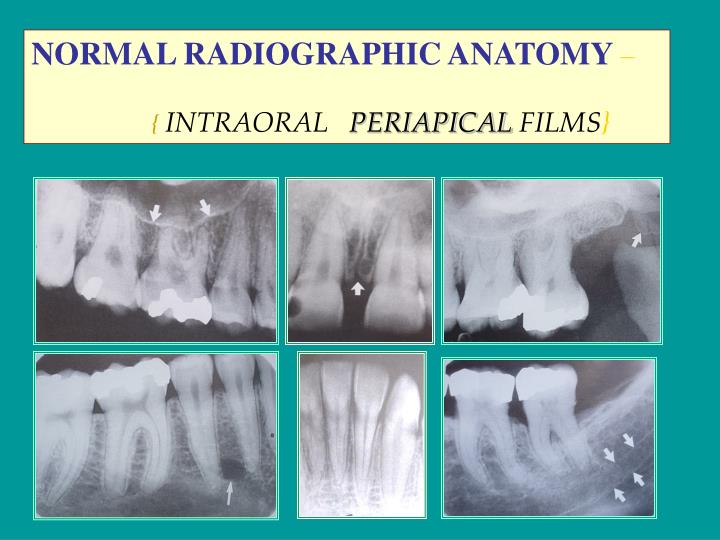
From thefuturedentistry.com
Anatomical Landmarks Focus Dentistry Maxillary Tuberosity And Hamular Notch The notch marks where the posterior border of the tuberosity (2) joins the medial (3) and lateral (4) plates of the pterygoid process. The hamular notch is a groove between the maxillary tuberosity and the hamular process; The hamular notch is a loose connective tissue, about 2 mm wide, located between the maxillary tuberosity and the. It is the posterior. Maxillary Tuberosity And Hamular Notch.
From www.pinterest.com
medialview of the maxilla Google Search Anatomia dos ossos Maxillary Tuberosity And Hamular Notch The hamular notch is critical to the design of the maxillary denture. The hamular notch is a loose connective tissue, about 2 mm wide, located between the maxillary tuberosity and the. The notch marks where the posterior border of the tuberosity (2) joins the medial (3) and lateral (4) plates of the pterygoid process. The maxillary wax rim can be. Maxillary Tuberosity And Hamular Notch.
From www.wikiwand.com
Palatine process of maxilla Wikiwand Maxillary Tuberosity And Hamular Notch Anatomical landmarks such as the retromolar pads, external oblique, mentalis muscle, frenum attachments, mylohyoid ridge, tuberosities, hamular notches, incisive papilla, labial sulcus, and buccal vestibule are critical and must be captured in the positive (and easily read in the negative) aspect of any edentulous impression. The notch marks where the posterior border of the tuberosity (2) joins the medial (3). Maxillary Tuberosity And Hamular Notch.
From www.joms.org
Maxillary Tuberosity Block Bone Graft Innovative Technique and Case Maxillary Tuberosity And Hamular Notch Anatomical landmarks such as the retromolar pads, external oblique, mentalis muscle, frenum attachments, mylohyoid ridge, tuberosities, hamular notches, incisive papilla, labial sulcus, and buccal vestibule are critical and must be captured in the positive (and easily read in the negative) aspect of any edentulous impression. The hamular notch is a loose connective tissue, about 2 mm wide, located between the. Maxillary Tuberosity And Hamular Notch.
From ar.inspiredpencil.com
Maxillary Tuberosity Maxillary Tuberosity And Hamular Notch Improper molding of this area could lead to soreness and loss of retention. The hamular notch is critical to the design of the maxillary denture. It is the posterior limit of the maxilla. Anatomical landmarks such as the retromolar pads, external oblique, mentalis muscle, frenum attachments, mylohyoid ridge, tuberosities, hamular notches, incisive papilla, labial sulcus, and buccal vestibule are critical. Maxillary Tuberosity And Hamular Notch.
From www.pinterest.com
Maxilla Bone Palatine Process; Alveolar Process Basic anatomy and Maxillary Tuberosity And Hamular Notch The hamular notch is a groove between the maxillary tuberosity and the hamular process; Anatomical landmarks such as the retromolar pads, external oblique, mentalis muscle, frenum attachments, mylohyoid ridge, tuberosities, hamular notches, incisive papilla, labial sulcus, and buccal vestibule are critical and must be captured in the positive (and easily read in the negative) aspect of any edentulous impression. The. Maxillary Tuberosity And Hamular Notch.
From www.slideserve.com
PPT NORMAL ANATOMICAL RADIOGRAPHIC LANDMARKS PowerPoint Presentation Maxillary Tuberosity And Hamular Notch The hamular notch is a loose connective tissue, about 2 mm wide, located between the maxillary tuberosity and the. It is the posterior limit of the maxilla. The hamular notch is critical to the design of the maxillary denture. Anatomical landmarks such as the retromolar pads, external oblique, mentalis muscle, frenum attachments, mylohyoid ridge, tuberosities, hamular notches, incisive papilla, labial. Maxillary Tuberosity And Hamular Notch.
From www.studocu.com
Landmarks for Complete Denture Anatomical landmarks and their Maxillary Tuberosity And Hamular Notch The hamular notch is a groove between the maxillary tuberosity and the hamular process; The hamular notch is critical to the design of the maxillary denture. Anatomical landmarks such as the retromolar pads, external oblique, mentalis muscle, frenum attachments, mylohyoid ridge, tuberosities, hamular notches, incisive papilla, labial sulcus, and buccal vestibule are critical and must be captured in the positive. Maxillary Tuberosity And Hamular Notch.
From exodontia.info
Fractured Maxillary Tuberosity Exodontia Maxillary Tuberosity And Hamular Notch The hamular notch is a groove between the maxillary tuberosity and the hamular process; The hamular notch is critical to the design of the maxillary denture. It is the posterior limit of the maxilla. The hamular notch is a loose connective tissue, about 2 mm wide, located between the maxillary tuberosity and the. The notch marks where the posterior border. Maxillary Tuberosity And Hamular Notch.
From thefuturedentistry.com
Anatomical Landmark of Mandible Focus Dentistry Maxillary Tuberosity And Hamular Notch Improper molding of this area could lead to soreness and loss of retention. The hamular notch is a groove between the maxillary tuberosity and the hamular process; The hamular notch is critical to the design of the maxillary denture. The notch marks where the posterior border of the tuberosity (2) joins the medial (3) and lateral (4) plates of the. Maxillary Tuberosity And Hamular Notch.
From prosthoguide.blogspot.com
Step by step Guide for Understanding the Prosthodontics Anatomical Maxillary Tuberosity And Hamular Notch The hamular notch is a loose connective tissue, about 2 mm wide, located between the maxillary tuberosity and the. Improper molding of this area could lead to soreness and loss of retention. Anatomical landmarks such as the retromolar pads, external oblique, mentalis muscle, frenum attachments, mylohyoid ridge, tuberosities, hamular notches, incisive papilla, labial sulcus, and buccal vestibule are critical and. Maxillary Tuberosity And Hamular Notch.
From exodontia.info
Fractured Maxillary Tuberosity Exodontia Maxillary Tuberosity And Hamular Notch The hamular notch is critical to the design of the maxillary denture. The notch marks where the posterior border of the tuberosity (2) joins the medial (3) and lateral (4) plates of the pterygoid process. It is the posterior limit of the maxilla. The hamular notch is a loose connective tissue, about 2 mm wide, located between the maxillary tuberosity. Maxillary Tuberosity And Hamular Notch.
From www.numerade.com
SOLVED Match the letter to its appropriate landmark Maxillary Maxillary Tuberosity And Hamular Notch Improper molding of this area could lead to soreness and loss of retention. The notch marks where the posterior border of the tuberosity (2) joins the medial (3) and lateral (4) plates of the pterygoid process. The hamular notch is a groove between the maxillary tuberosity and the hamular process; It is the posterior limit of the maxilla. The hamular. Maxillary Tuberosity And Hamular Notch.
From pocketdentistry.com
7 Skull and oral anatomy Pocket Dentistry Maxillary Tuberosity And Hamular Notch It is the posterior limit of the maxilla. Improper molding of this area could lead to soreness and loss of retention. The hamular notch is a loose connective tissue, about 2 mm wide, located between the maxillary tuberosity and the. The hamular notch is critical to the design of the maxillary denture. The notch marks where the posterior border of. Maxillary Tuberosity And Hamular Notch.
From www.animalia-life.club
Zygomatic Bone Maxillary Process Maxillary Tuberosity And Hamular Notch The hamular notch is critical to the design of the maxillary denture. The notch marks where the posterior border of the tuberosity (2) joins the medial (3) and lateral (4) plates of the pterygoid process. Anatomical landmarks such as the retromolar pads, external oblique, mentalis muscle, frenum attachments, mylohyoid ridge, tuberosities, hamular notches, incisive papilla, labial sulcus, and buccal vestibule. Maxillary Tuberosity And Hamular Notch.
From quizlet.com
MAXILLARY LANDMARKS Diagram Quizlet Maxillary Tuberosity And Hamular Notch Anatomical landmarks such as the retromolar pads, external oblique, mentalis muscle, frenum attachments, mylohyoid ridge, tuberosities, hamular notches, incisive papilla, labial sulcus, and buccal vestibule are critical and must be captured in the positive (and easily read in the negative) aspect of any edentulous impression. The hamular notch is a groove between the maxillary tuberosity and the hamular process; The. Maxillary Tuberosity And Hamular Notch.
From internationalclinics.dental
Maxillary Orthognathic Surgery International Clinics Maxillary Tuberosity And Hamular Notch It is the posterior limit of the maxilla. Improper molding of this area could lead to soreness and loss of retention. The notch marks where the posterior border of the tuberosity (2) joins the medial (3) and lateral (4) plates of the pterygoid process. The hamular notch is a groove between the maxillary tuberosity and the hamular process; The maxillary. Maxillary Tuberosity And Hamular Notch.